Featured · TodayScience
Optogenetics: Controlling Brain Cells With Light
How a light-sensitive protein from pond algae gave neuroscientists a switch for turning specific brain cells on and off with pulses of blue light.
Listen
Read by Finch · 8 min
A pond organism learns to steer neurons
The protein that remade modern neuroscience spends most of its working life helping a single-celled green alga decide which way to swim.
Chlamydomonas reinhardtii uses a tiny eyespot to sense light and a pair of flagella to swim toward or away from it, a behavior called phototaxis. Researchers chasing the mechanism behind that reflex eventually traced it to two rhodopsin genes in the algal genome, proteins related to the light receptors in human eyes but wired very differently3.
The real groundwork went back further. In 1971, Dieter Oesterhelt and Walt Stoeckenius described bacteriorhodopsin, a light-driven proton pump in a salt-loving archaeon, proving that microbes could turn photons directly into ion movement6. That observation sat in the literature for decades as a curiosity in membrane biophysics.
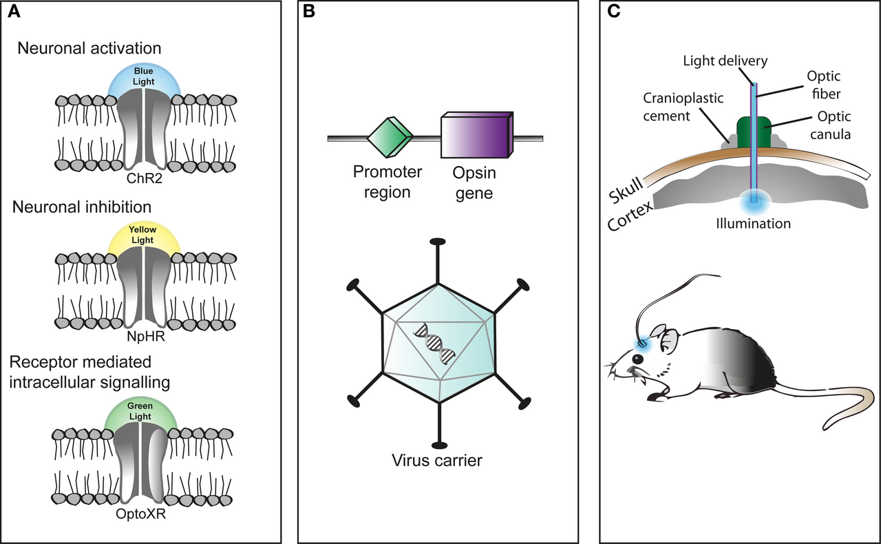
The breakthrough with the algal genes came in 2002 and 2003, when Georg Nagel, Ernst Bamberg, Peter Hegemann, and colleagues expressed them in frog eggs and found something unusual: unlike animal rhodopsins, which trigger a slow cascade of internal signaling, these proteins were themselves light-gated channels. They named them channelrhodopsin-1 and channelrhodopsin-2 (ChR2), and showed that blue light caused ChR2 to let positive ions flood into a cell almost instantly2,7. In their 2003 paper, they floated the idea that this might be useful for controlling membrane voltage in mammalian cells, but at that point it was still an algal curiosity. Nobody had tried it in a working neuron2.
That changed in March 2004, when Karl Deisseroth, a psychiatrist and neuroscientist who had just started his lab at Stanford, read the channelrhodopsin papers and emailed Nagel asking for the gene3. Nagel sent the DNA, and Ed Boyden, then a graduate student in Deisseroth's lab, packaged it into a lentivirus and infected cultured rat hippocampal neurons with it1,4. Accounts differ on the exact date of the first clean result, with Deisseroth's lab notebook pointing to early July 2004 and other retellings citing an August 2004 experiment, but the gist is the same: shine blue light on a ChR2-loaded neuron, and it fires1,3.
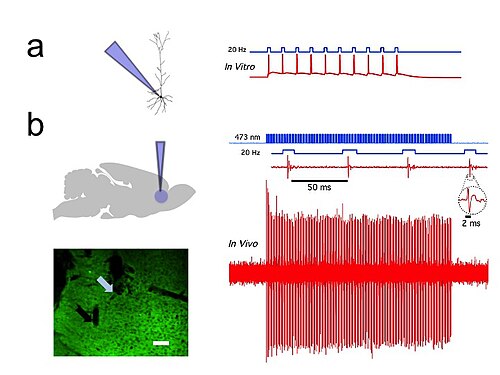
The full demonstration landed in August 2005, when Boyden, Feng Zhang, Bamberg, Nagel, and Deisseroth published "Millisecond-timescale, genetically targeted optical control of neural activity" in Nature Neuroscience. The paper showed that ChR2-expressing hippocampal neurons produced strong photocurrents, fired action potentials on command, tracked rapid pulses of light with millisecond fidelity, and did all this without obvious harm to the cells under the conditions tested1,4. What made the paper matter wasn't just that ChR2 responded to light. It was that Deisseroth's team had assembled a complete toolkit, gene, delivery method, light source, and electrical proof, into a single working system that any lab could, in principle, copy4,8.
Deisseroth gave the resulting field a name the following year: optogenetics, a fusion word for a method that is exactly as literal as it sounds5,4.
Not a one-lab story
The Stanford result landed first in the public imagination, but it was not the only group circling the same idea in 2005 and 2006.
Alexander Gottschalk's team got channelrhodopsin working in the roundworm Caenorhabditis elegans, controlling the animal's behavior with light. Stefan Herlitze's group and Hiromu Yawo's group separately demonstrated optical control of excitable cells using related channelrhodopsin tools. And Zhuohua Pan, working on restoring vision to blind mice, used ChR2-related methods in retinal tissue, an early hint at the therapeutic path optogenetics would eventually take2.
These parallel efforts mattered because they showed the method wasn't a fluke of one cell type or one species. ChR2 worked in worm neurons, mouse brain slices, chicken embryos, and cultured cell lines, which told researchers they had found something closer to a general-purpose tool than a lab trick2.
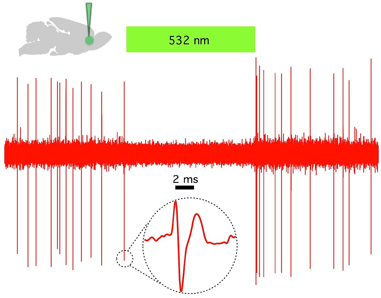
The toolkit also grew quickly on the inhibitory side. Within a couple of years, Deisseroth's and Boyden's labs, working alongside Gottschalk and Nagel, added halorhodopsin and other opsins that could silence neurons with a different color of light, giving researchers an on switch and an off switch they could use in the same experiment1,4.
Who's who
- Karl Deisseroth: Stanford psychiatrist and bioengineer who requested the channelrhodopsin gene in 2004, led the 2005 demonstration, and coined the term "optogenetics" in 20063,4.
- Ed Boyden: Graduate student in Deisseroth's lab who adapted ChR2 for neurons using a lentiviral vector and was lead author on the 2005 paper1,4.
- Feng Zhang: Graduate student in Deisseroth's lab, co-author on the 2005 paper1,4.
- Georg Nagel and Ernst Bamberg: Max Planck Institute of Biophysics researchers who characterized ChR2 as a light-gated channel in 2002-2003 and supplied the gene to Deisseroth2,7,3.
- Peter Hegemann: University of Regensburg biophysicist who studied algal phototaxis and co-discovered the fast photocurrents that led to channelrhodopsin3.
- Gero Miesenböck: Reported an earlier genetically targeted light-control method using Drosophila rhodopsin in cultured mammalian neurons in January 2002, a precursor approach that predates channelrhodopsin-based optogenetics1.
- Zhuohua Pan: Wayne State University researcher who applied channelrhodopsin to retinal cells in pursuit of vision restoration2.
Who got there first, and when
The tidiest version of this story credits Deisseroth's Stanford lab with inventing optogenetics outright in 2005. The real picture is messier, and sources disagree on some of the specifics.
There is a dating dispute inside Deisseroth's own lab records: some accounts point to a notebook entry from early July 2004 as the first demonstration of light-driven neuronal firing, while other retellings describe the decisive experiment as happening in August 20041,3. Which one counts as the "true" first depends on how strict a standard you apply.
Zhuohua Pan has stated that he observed optical activation of retinal neurons with channelrhodopsin as early as February 2004, which would put him roughly five months ahead of Deisseroth's group, though Pan's full in vivo results were not published until 20051. Deisseroth's team, by contrast, published the first complete demonstration in a mainstream mammalian neuron system with the methodological package (viral delivery, defined light source, electrophysiological proof) that became the field's template1,4.
So the honest framing is less "who invented it" and more "who assembled the parts into something reproducible first," and on that score Deisseroth's 2005 paper is the one that the field converged on as its founding document, even though the underlying components, the gene, the biophysics, the viral tools, came from several labs working somewhat independently4,8.
Timeline
- 1971Dieter Oesterhelt and Walt Stoeckenius describe bacteriorhodopsin, a light-driven proton pump in an archaeon.
- 2002-2003Georg Nagel, Ernst Bamberg, and Peter Hegemann characterize channelrhodopsin-1 and channelrhodopsin-2 as light-gated ion channels.
- March 2004Karl Deisseroth contacts Nagel requesting the ChR2 gene for use in neurons.
- July-August 2004Deisseroth's lab records the first light-driven firing of a ChR2-expressing neuron; exact date is disputed.
- August 2005Boyden, Zhang, Bamberg, Nagel, and Deisseroth publish the landmark Nature Neuroscience paper demonstrating millisecond optical control of neurons.
- 2006Deisseroth coins the term 'optogenetics.'
Pictures


Sources
- OptogeneticsWikipedia
- He may have invented one of neuroscience's biggest ...statnews.com
- From channelrhodopsins to optogenetics - EMBO Molecular Medicinelink.springer.com · 2013
- Light activated - NatureNature · 2008
- Two Decades of Optogenetic Tools: A Retrospective and a Look AheadPubMed Central · 2025
- Optogenetics: shining a light on the brain | Bioscience ...academic.oup.com
- A history of optogenetics: the development of tools for controlling brain circuits with lightNCBI · 2011
- The optogenetic (r)evolution - Molecular Genetics and Genomicslink.springer.com · 2011
- Karl DeisserothWikipedia
- Generative OptogeneticsHacker News · 2025
- Augmenting biological intelligence with RL in C.elegans using optogenetics [pdf]Hacker News · 2024
Researched and written by Lorelet from the sources above. It cites as it goes, but it can still get things wrong, so check the sources for anything that matters.
Report a problem
More featured lorelets
See all
Today · Technology
ChatGPT Started Forging New Yorker Cartoonists' Signatures
When an AI model began signing fake New Yorker cartoons with real cartoonists' names, it turned a style-imitation problem into something that felt, t…

Today · Music
The Monkees' Missing Tapes: Theft, Rescue, or Both?
Somewhere between corporate indifference and collector obsession, the Monkees' original session tapes went missing for decades. The story of how they…

Oct 5 · History
Eugene Hasenfus: The Crash That Exposed Iran-Contra
A cargo kicker from Wisconsin parachuted into the Nicaraguan jungle with a weapons flight behind him and, in trying to explain his job, blew open one…